Maxillary Sinus Lift for Dental Implants: When It Is Recommended and How It Is Performed
A maxillary sinus lift, also known as a sinus graft, is a procedure used for bone grafting when there is insufficient bone in the posterior region of the upper jaw for dental implants. The goal of dental bone grafting is to increase bone height to allow for the placement of dental implants. The procedure is performed through a small incision in the gum on the side of the bone defect.
This situation is relatively common because the upper posterior teeth are located near the maxillary sinus, a cavity inside the upper jaw. After tooth loss, bone resorption and expansion of the maxillary sinus may occur progressively, reducing the amount of bone available to support implants.
When there is insufficient bone height, a sinus lift may be recommended to increase bone volume in the area and create more favorable conditions for the placement of dental implants.
At the ImplArt Clinic in São Paulo, the decision to perform a bone graft is made after a clinical evaluation and analysis of imaging tests—particularly CT scans—by Dr. Roberto Markarian. This analysis allows for a detailed examination of the anatomy of the maxillary sinus and the available bone volume.
Does every patient with insufficient bone need a zygomatic implant?
According to Dr. Roberto Markarian, PhD, Clinical Director of ImplArt Clinic, no. “A lack of bone, in and of itself, does not mean that the patient needs zygomatic implants. In many cases, we can rebuild bone volume using grafting techniques and create the conditions necessary to use conventional dental implants.”
The zygomatic implant is an important alternative for selected cases of severe maxillary atrophy, but it uses longer implants anchored in the zygomatic bone and requires careful consideration of the indication. For this reason, sinus grafting is an extremely important tool for rehabilitating the patient and creating the necessary bone height for the placement of conventional implants.
According to Dr. Roberto: “When it is still possible to reconstruct the area and use conventional implants, this option should be considered. The goal is to choose the most appropriate solution for each patient, while also keeping in mind the final outcome of the rehabilitation.”
At ImplArt Clinic, this planning can be part of high-quality restorative treatments, including zirconia protocol prostheses—a premium option that combines strength, high quality, durability, and the potential for a more natural-looking result.
Watch the video below, where we discuss bone grafting techniques for dental implants in more detail:

What is a maxillary sinus lift?
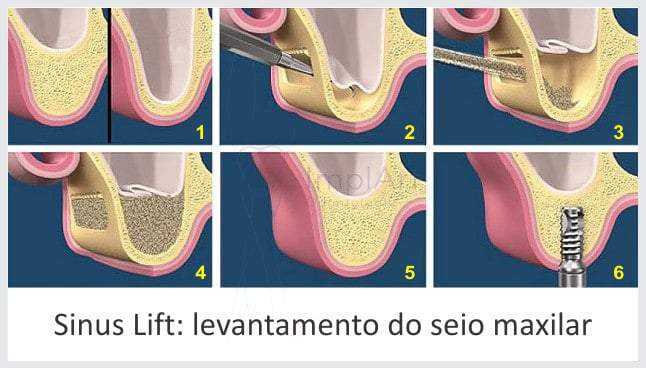
A sinus lift is a surgical procedure performed in the posterior region of the maxilla to create space for the formation of new bone tissue beneath the maxillary sinus.
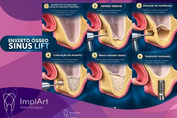
In the lateral window technique, a small opening is created in the lateral wall of the maxilla. Through this opening, the membrane that lines the interior of the maxillary sinus—known as the sinus membrane or Schneider’s membrane—is carefully elevated.
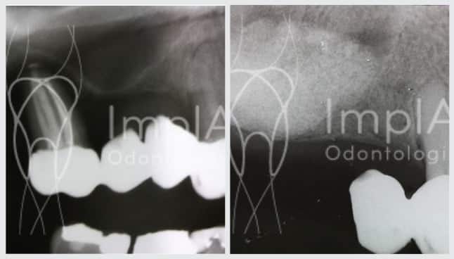
A variable amount of bone regeneration precursor material is placed inside this opening. As the healing process progresses, the area regenerates, increasing the amount of mineralized tissue available for dental implant planning.
After sinus lift surgery, you should wait 6 months for the area to regenerate before having the implants placed.
Watch the video below, where we discuss maxillary SINUS LIFT GRAFTING in more detail: When There Isn’t Enough Bone for a Dental Implant:

Why might there not be enough bone to place implants in the posterior region of the maxilla?
Bone loss can occur for various reasons, but one of the most common causes is the loss or extraction of teeth.
The bone surrounding the tooth roots exists, in part, to provide support for the teeth. When a tooth is lost and not replaced, this region no longer receives the same functional stimuli and may undergo progressive resorption.
In the posterior region of the maxilla, there is yet another important phenomenon: the maxillary sinus may expand into the area previously occupied by bone and tooth roots.
When bone resorption of the alveolar ridge is combined with maxillary sinus expansion, the distance between the mouth and the floor of the sinus can become very small.
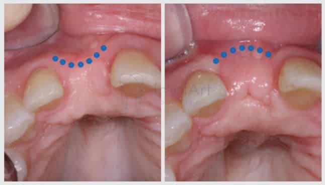
For this reason, some patients have adequate bone width but insufficient bone height for the safe placement of a dental implant.
Materials Used in Grafting
Nowadays, there are a variety of materials that can be used for maxillary sinus grafting:
- Freeze-dried bovine bone – Genox
- Autogenous bone (from the person’s own body)
- Synthetic hydroxyapatite (bone precursor)
- Allogeneic bone from a bone bank (bone graft)
- Synthetic Glass Materials – Bioss
Studies show that all materials are suitable for this type of graft; the choice depends primarily on the surgeon’s preference.
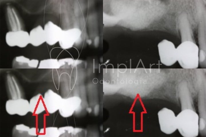
Bone loss: causes
Bone loss can occur for a number of reasons, but the most common is tooth loss or extraction without immediate replacement. When a person loses teeth, the body resorbs the bone in the upper or lower jaw because it perceives that the bone no longer serves a function (which was to support the teeth). This loss is progressive, and over the years, the person’s bone volume decreases in both height and width, which initially makes it difficult to place dental implants. If you have any questions, please contact us or schedule an appointment with our team.
When is a maxillary sinus graft recommended?
A sinus lift may be indicated when there is insufficient bone below the maxillary sinus to accommodate the planned implant.
This situation may be related to:
- loss of upper posterior teeth many years ago;
- bone resorption following tooth extractions;
- expansion or pneumatization of the maxillary sinus;
- previous periodontal disease;
- bone loss resulting from dental infections;
- the patient’s individual anatomical characteristics.
However, having insufficient bone does not automatically mean that every patient will need a sinus lift.
The indication depends on the quantity and quality of the residual bone, the anatomy of the maxillary sinus, the planned implant position, and the specific characteristics of each case.
How do I know if I need a sinus lift?
Diagnosis begins with a clinical evaluation and imaging tests.
A panoramic X-ray can provide an initial view of the area, but a CT scan allows for a much more detailed three-dimensional analysis.
CT scans can be used to evaluate, among other factors:
- the height of the existing bone;
- the thickness of the bone crest;
- the shape of the maxillary sinus;
- the thickness of the side wall;
- the presence of bony septa;
- the characteristics of the sinus membrane;
- the planned position of the implants;
- anatomical relationships important for surgery.
This information makes it possible to develop a more personalized treatment plan and determine whether a graft is truly necessary and, if so, how large it should be.
How is a sinus lift performed?
In the lateral window sinus lift technique, the procedure can be broken down into a few main steps.
1. Assessment of the region
First, clinical and imaging tests are analyzed to determine the amount of available bone and the anatomy of the maxillary sinus.
2. Access to the lateral wall of the maxilla
During the surgery, an incision is made in the gum to expose the lateral wall of the maxillary bone.
3. Creation of the bone window
A small window is created in the lateral wall of the maxilla to allow access to the interior of the sinus region.
4. Maxillary sinus membrane elevation
The sinus membrane is carefully separated from the floor of the sinus and elevated.
This is one of the most delicate steps in the procedure.
5. Placement of the grafting material
The space created beneath the elevated membrane is filled with the biomaterial selected for the case.
This material supports the process of bone regeneration and formation.
6. Closure and healing period
After the area has been filled, the surgical incision is closed, and the healing process begins.
Over the following months, the biological process of graft integration and remodeling takes place.
What materials can be used for a maxillary sinus graft?
There are various biomaterials that can be used in bone regeneration procedures.
Among them are:
Autogenous graft
It is bone obtained from the patient himself.
Depending on the clinical situation, it can be used alone or in combination with other biomaterials.
Biomaterial of bovine origin
Xenogeneic bone grafts derived from cattle are widely used in bone regeneration procedures and can serve as a scaffold for the formation and maintenance of new bone mass.
Allogeneic graft
It is derived from human bone tissue that has been processed and made available by authorized tissue banks.
Synthetic biomaterials
There are also synthetically produced materials, such as certain calcium phosphates and hydroxyapatites.
The choice of material does not depend solely on the practitioner’s preference. Factors such as the required volume, the characteristics of the defect, the anatomy of the region, the surgical technique used, and the treatment plan involving implants are all taken into account.
In certain situations, different materials or combinations of materials may be used.
Is it possible to place the implant at the same time as the sinus lift?
In some cases, yes.
Maxillary sinus graft surgery and implant placement can be performed during the same procedure when there is sufficient quantity and quality of residual bone to provide adequate stability for the implant.
However, when there is little remaining bone structure or when it is not possible to achieve the necessary stability, a two-stage treatment approach may be more appropriate.
In this case, the sinus lift is performed first. After the bone regeneration period, a new evaluation is conducted to plan and place the implants.
Therefore, there is no single sequence that is valid for all patients.
The decision must be made on a case-by-case basis.
How long does it take for a maxillary sinus graft to heal?
Bone regeneration is a biological process and takes time.
The healing period may vary depending on:
- initial amount of bone present;
- graft volume;
- biomaterial used;
- the patient’s individual characteristics;
- duration of the procedure;
- whether or not the implant can be placed at the same time.
Therefore, it is not appropriate to set a single deadline for all cases.
Throughout treatment, imaging tests may be used to monitor the progress of the grafted area before continuing rehabilitation with implants.
Is a sinus lift a safe procedure?
The maxillary sinus lift is a well-established technique in implant dentistry that has been used for decades to allow for the placement of implants in posterior regions of the maxilla with bone deficiency.
Like any surgery, however, it requires diagnosis, planning, and a thorough understanding of the anatomy of the area.
One of the most common complications is perforation of the maxillary sinus membrane during its elevation.
The presence of bony septa, membrane characteristics, and anatomical variations can make certain cases more complex.
When a perforation occurs, how it is managed depends on its characteristics and extent. In many cases, there are specific techniques for treating it during the procedure itself.
Therefore, a preliminary assessment of the anatomy using CT imaging is an important step in the planning process.
Can people with sinusitis undergo a sinus lift?
Any abnormalities in the maxillary sinus must be evaluated before surgery.
Patients with symptoms or signs of sinus disease may require further testing and, in certain situations, a medical or otolaryngological evaluation prior to the procedure.
This does not mean that a person who has previously had sinusitis is automatically unable to undergo a maxillary sinus graft.
The recommendation depends on the current condition of the maxillary sinus and an individual assessment of the case.
Can a maxillary sinus graft change the shape of the face?
Under normal circumstances, that is neither the goal nor the expected outcome of a sinus lift.
The graft is performed internally, in the maxillary sinus region, to create bone volume for implant placement.
Therefore, the procedure is not performed to augment the cheekbones or alter the outer contours of the face.
Can a sinus lift affect breathing?
The maxillary sinus graft is performed in an anatomical region separate from the main air passage through the nasal cavities and is not intended to block the airways.
However, patients who already have shortness of breath, sinus problems, or otolaryngological symptoms should report these conditions during the evaluation.
Each case must be evaluated individually before surgery.
What happens after sinus lift surgery?
In the first few days, a recovery period similar to that of other dental surgical procedures is expected.
Postoperative instructions vary depending on each case and must be followed strictly.
In general, it is important to avoid situations that cause excessive pressure to build up inside the maxillary sinus during the initial healing period.
The patient receives specific instructions regarding hygiene, diet, and medications when prescribed, as well as instructions for follow-up visits.
What precautions are important to take after a sinus lift?
The recommendations depend on the type of surgery performed, but may include measures such as:
- take the prescribed medication as directed;
- avoid physical exertion as directed by a healthcare professional;
- Do not touch the operated area;
- maintain hygiene in accordance with the instructions received;
- avoid increased pressure in the maxillary sinus area during the first few days;
- attend follow-up appointments.
The postoperative period should be monitored by the healthcare professional responsible for the treatment.
Is a sinus lift always necessary when there is a lack of bone?
No.
Currently, there are various strategies for treating patients with bone loss in the posterior region of the maxilla.
The choice depends on the amount of available bone, the patient’s anatomy, the position of the future teeth, the characteristics of the planned implants, and the goals of the rehabilitation.
Therefore, CT scans and individualized planning are essential before recommending any grafting technique.
Sinus Lift and Implant Planning at ImplArt Clinic
At the ImplArt Clinic in São Paulo, dental implant and bone graft treatments are planned on an individualized basis.
The evaluation may include X-rays and CT scans, digital planning, and modern implant dentistry techniques, allowing for a preliminary assessment of the region’s anatomy and the planned position of the implants.
The goal of planning is not simply to determine whether or not bone is present, but to identify the best strategy for achieving functional and predictable rehabilitation for each patient.
Dr. Roberto Markarian, Ph.D.
Dr. Roberto Markarian is a specialist in Implant Dentistry and Prosthodontics, holds a master’s degree and a Ph.D., and serves as Clinical Director of the ImplArt Clinic.
He has extensive experience in implant dentistry and oral rehabilitation, having placed more than 12,000 dental implants, in addition to his academic and scientific work.
He is the author and co-author of international scientific chapters on digital implantology, computer-guided surgery, and artificial intelligence applied to dental prosthetics, published by publishers such as Wiley and Springer Nature.
CRO-SP 73,583.
Frequently Asked Questions About Sinus Lifts
What does “sinus lift” mean?
A sinus lift refers to the elevation of the maxillary sinus. It is a technique used to increase bone availability below the maxillary sinus when there is insufficient bone height for the placement of dental implants.
What is the purpose of a maxillary sinus graft?
Its main purpose is to promote bone augmentation in the posterior region of the maxilla in order to enable or improve the conditions for placing dental implants.
Does every patient who has lost upper molars need a sinus lift?
No. The need depends on the amount of remaining bone and the individual anatomy of the region.
Can the implant be placed at the same time as the graft?
In certain cases, yes. This depends mainly on whether adequate stability of the implant can be achieved in the existing bone.
How long after a sinus lift can an implant be placed?
The duration varies depending on the characteristics of the case, the volume of graft material, the material used, and the quality of the residual bone. In some patients, the implant can be placed during the same procedure; in others, it is necessary to wait for bone regeneration.
Does a sinus lift hurt?
The procedure is performed under anesthesia. During the postoperative period, discomfort, swelling, and tenderness may occur to varying degrees; these symptoms should be monitored and managed according to professional guidance.
Can people with sinusitis undergo a maxillary sinus graft?
It depends on the current condition of the maxillary sinus. Any sinus abnormalities must be identified and evaluated before surgery.
Can a facial implant change the shape of the face?
The procedure is performed in-office and is not intended to alter the external appearance of the face.
Do I need to have a CT scan before a sinus lift?
Computed tomography is a highly important imaging test for three-dimensional assessment of bone volume and the anatomy of the maxillary sinus prior to surgical planning.
Evaluation for Maxillary Sinus Grafting and Dental Implants
The need for a sinus lift can only be determined following an individualized evaluation by our clinical director, Dr. Roberto Markarian.
At ImplArt Clinic, the treatment plan developed by Dr. Markarian takes into account the patient’s clinical condition, imaging studies, the amount of available bone, and the type of restoration desired.
To schedule a consultation with the team at Clínica ImplArt in São Paulo, please contact us:
WhatsApp / Phone: (11) 3262-4750
Learn more about the treatments offered at Clinica ImplArt
To learn more about the treatments offered at ImplArt Dental Clinic, check out photos of our treatments on Instagram at https://www.instagram.com/clinicaimplart/
Our videos on YouTube: https://www.youtube.com/@ImplArtImplanteseestetica
Check out our Pinterest page: https://br.pinterest.com/implante/
To view our photo gallery of our treatments, click here!
Contact us on WhatsApp at (11) 3262-4750
Technical article by DR. ROBERTO MARKARIAN – CRO-SP 73.583
, Founder and Director of the ImplArt Dental Clinic. – Dr. Roberto’s LinkedIn profile
Dr. Roberto Markarian is a renowned implant dentist practicing in São Paulo. He is a leading authority on dental implants and computer-aided dental prosthetics in Brazil. In addition to having experience with more than 12,000 implants placed, he is a researcher who produces knowledge that reaches a global audience through publications in renowned scientific journals in the field of dentistry. He is responsible for promoting knowledge and cutting-edge technology applied to all treatments offered by the ImplArt Dental Clinic.
Dr. Markarian has just contributed to an internationally renowned publication featuring cutting-edge research on the use of artificial intelligence in the production of dental prostheses, in the chapter “Artificial Intelligence in Prosthodontics,” published by Springer Nature / Springer, Cham, in the book *Dental Clinical Procedures using Artificial Intelligence*.
 Agende sua consulta agora por WhatsApp