

Can People with Dental Implants Undergo an MRI? Differences Between Titanium and Zirconia Dental Implants
This is a common question we receive from our patients who have dental implants and have been referred by their doctor for an MRI. After all, can people with dental implants undergo an MRI?
In most cases, the answer is yes. People with titanium or zirconia dental implants can usually undergo an MRI.
According to Dr. Roberto Markarian, director of the ImplArt Clinic, the most important precaution is to inform the doctor and the team conducting the exam in advance that you have implants, prostheses, or other devices in your body.
But there is an important difference that is not always explained to the patient: it is one thing to ensure it is safe to undergo an MRI; it is quite another to consider the possibility that dental materials might interfere with the quality of the images obtained.
This distinction is especially important when the MRI involves the head, face, mandible, maxilla, or structures near the mouth. But don’t worry, we’ll explain everything in detail below!

Why are there concerns about dental implants and MRI scans?
Magnetic resonance imaging (MRI) uses a strong magnetic field to produce detailed images of the body. For this reason, before the exam, patients are asked about any implants, prostheses, medical devices, or metallic materials they may have.
This causes many people with dental implants to worry. Modern dental implants are primarily made of titanium or zirconia, materials that have been used for many years in implant dentistry.
Having a dental implant, therefore, does not automatically mean that an MRI is contraindicated. What needs to be considered is the type of material present, the area to be examined, and whether there is a possibility of image interference.
Security and image interference are not the same thing
This is probably the most important point for understanding the subject.
According to Dr. Markarian, an implant may be compatible with an MRI scan and still cause some degree of distortion in the image. These distortions are called image artifacts.
In practice, this means that certain structures near the implant may appear distorted, with areas of signal loss or reduced clarity. This tends to be more significant when the doctor needs to examine an area near the mouth.
In a knee MRI, for example, a dental implant is very far from the area being examined. However, in certain exams of the face, jaw, or head, the presence of dental materials may have a greater impact on image quality. In other words, according to Dr. Markarian, the interference is limited to the area near the implant and does not affect the exam as a whole.
Can people with titanium dental implants have an MRI?
Titanium dental implants are the most commonly used in implant dentistry and, in general, do not prevent patients from undergoing an MRI.
Titanium has properties that are quite different from those of strongly ferromagnetic materials, which often raise concerns during screening for an MRI.
On the other hand, because it is a metallic material, titanium can cause some degree of image artifact, especially when the exam involves areas near the implant.
Studies that have analyzed dental implants using magnetic resonance imaging show that the intensity of these artifacts varies depending on the material, the sequence used by the equipment, and the region being examined.
Therefore, it is not accurate to simply say that “metal cannot be brought into the MRI scanner.” There are many types of metallic materials and medical devices, each with its own characteristics. The assessment must be made by the team responsible for the exam.
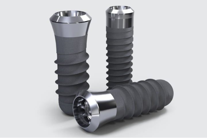
And can people with zirconia implants have an MRI?
Zirconia dental implants are ceramic and behave differently from metal implants during imaging.
Comparative studies of titanium and zirconia implants indicate that ceramic implants may produce fewer artifacts in certain MRI sequences.
This can make it easier to visualize the structures surrounding the implant in certain types of imaging studies. However, this characteristic does not mean that zirconia is automatically the best implant option for all patients.
The choice between a titanium implant and a zirconia implant should take into account the overall treatment plan: bone quantity and quality, implant position, gum tissue, prosthesis characteristics, aesthetics, masticatory function, and the patient’s individual condition.
The possibility of undergoing future MRI scans is just one of many factors that can be taken into account.
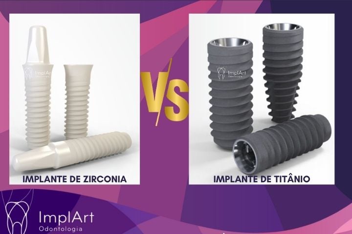
Differences between titanium dental implants and zirconia implants for an MRI
| Feature | Titanium Implants | Zirconia Implants |
|---|---|---|
| Material Type | Non-ferromagnetic metal (paramagnetic) | Non-magnetic ceramic (metal-free) |
| Patient Safety | 100% Safe. Will not pull, shift, or heat up. | 100% Safe. Will not pull, shift, or heat up. |
| MRI Image Artifacts | Causes moderate image distortion (dark shadows/signal voids) directly around the implant area. | Causes minimal to no distortion. The surrounding tissue remains clearly visible. |
| Impact on Diagnosis | May slightly interfere with clear views of the jaw, tongue, or sinuses if the scan is focused on the face. | Excellent for head and neck imaging; allows clear depiction of the bone-to-implant interface. |
Magnetic Interaction & Composition: Titanium is a biocompatible metal. While it is technically paramagnetic, it is non-ferromagnetic, meaning it does not react strongly to the MRI magnet. Zirconia, on the other hand, is a completely metal-free ceramic, making it entirely non-magnetic
Image Artifacts (The Main Difference): During a brain, head, or neck MRI, titanium implants cause a “susceptibility artifact”. This shows up as a small dark shadow or distortion on the scan, making the immediate surrounding tissues harder to see. Zirconia implants produce virtually no distortion, allowing radiologists to get a perfectly clear picture of the jawbone and surrounding anatomy.
Can the implant be attracted to the MRI machine?
This concern is understandable because the MRI machine produces a very strong magnetic field.
A conventional dental implant made of titanium or zirconia, once installed and osseointegrated, does not behave like a loose metal object placed inside the device. Therefore, the examination does not exert enough force to dislodge the osseointegrated dental implant from its position.
Even so, Dr. Roberto Markarian, an implantologist and director of the ImplArt Clinic, advises that it is essential to disclose the presence of any implants before the exam.
The screening performed by the radiology department is designed precisely to enable the team to identify all devices present in the body and determine whether any specific precautions are necessary.
Do dental implants interfere with brain or head MRI scans?
Some degree of interference may occur, depending mainly on the location of the implant and the type of exam performed. When the purpose of the MRI is to evaluate structures near the oral cavity, mandible, maxilla, or certain regions of the face, the artifacts produced by dental materials may be more significant.
The farther the examined region is from the mouth, the less direct influence the implant tends to have on the area of interest.
According to Dr. Markarian, it is important to note that image artifacts do not necessarily pose a risk to the patient. In some situations, the challenge lies simply in obtaining a sufficiently clear image of the region that needs to be evaluated.
The radiology team can take this information into account when determining the exam protocol.
Can people with dental implants have an MRI?
Patients with implant-supported prostheses can also undergo MRI scans in many situations.
However, a prosthetic restoration is a more complex assembly than a single implant.
It may involve:
- dental implants;
- prosthetic intermediaries;
- screws;
- metal components;
- internal structure of the prosthesis;
- ceramic or resin-based materials.
For this reason, it is important to inform the radiology staff not only that the patient has implants, but also that the patient wears a fixed prosthesis over them.
According to Dr. Markarian, if there are any questions about the materials used in the restoration, the dentist responsible for the treatment can provide additional information. In addition, he notes that there are now modern materials, such as zirconia, that allow for the creation of metal-free dental prostheses. This material can be an alternative for patients who do not want metal in their dental prostheses.
Can crowns and other dental prosthetics also interfere?
Depending on the material used, yes. Today, there are crowns and dentures made from different materials, such as:
- zirconia;
- ceramics;
- resin;
- titanium;
- metal alloys;
- combinations of different materials.
Each material may behave differently during image acquisition.
Therefore, when a patient has undergone extensive dental rehabilitation, has a protocol prosthesis, an overdenture, or special prosthetic components, they should share information about their treatment with the team responsible for the MRI.
According to Dr. Roberto Markarian, modern materials such as zirconia are the first choice for many patients. Currently, many patients prefer these more modern materials because they offer a number of benefits: excellent aesthetics, durability, and the absence of metal.
I need to know the brand of my implant.
In most cases, the first step is simply to mention that a dental implant is present. However, in specific situations, the radiology team may request additional information about the device.
For this reason, it is advisable to keep records related to dental treatment, especially when more complex procedures have been performed. When necessary, the dental practice or the dentist in charge can help identify the implant system used and the materials in the prosthesis.
Can a magnetic field dislodge a dental implant?
According to Dr. Roberto Markarian, this is also a concern that some patients express before the procedure, but he explains that an osseointegrated dental implant is integrated into the bone tissue. It is not simply supported or superficially embedded in the mouth; therefore, removing a dental implant requires a great deal of physical force.
With conventional dental implants made of titanium or zirconia, the most frequently discussed issue regarding MRI scans relates to the formation of artifacts and image quality, rather than implant loosening caused by the device.
Even so, any patient with implants or medical devices must follow the safety protocol of the facility where the exam will be performed.
What should I let them know before having an MRI?
When you schedule your appointment or arrive for your exam, please mention that you have dental implants.
If you know, please also provide the following information:
- whether the implants are made of titanium or zirconia;
- if you have a protocol prosthesis;
- if an overdenture is used;
- whether the prosthesis contains any metal components;
- if you have other devices implanted in your body.
If more specific information is needed, the radiology team may request it. In case of uncertainty, the dentist in charge of the treatment can assist by providing information about the system used.
The decision regarding the conditions for performing the MRI and the appropriate protocol rests with the physician and the radiology team responsible for the exam.
So, can people with dental implants have an MRI?
Most of the time, yes.
Having a titanium or zirconia dental implant does not usually prevent you from undergoing an MRI. The most important precaution is to inform the staff who will be performing the exam in advance that you have implants and any dental prostheses.
It is also important to distinguish between two issues: safety during the MRI scan and the possibility of image interference.
Dental materials can cause artifacts, especially when the area being examined is close to the mouth. This does not, however, automatically mean that the exam is contraindicated.
Titanium or zirconia: Is there a difference on an MRI?
The main difference lies in the formation of artifacts.
Studies comparing titanium dental implants with zirconia ceramic implants found that zirconia implants caused less image artifacts under certain examination conditions.
From the perspective of a patient who already has titanium implants, this does not mean that there is an indication to replace those implants with zirconia implants. They are different materials, each with its own characteristics and indications.
The decision regarding which implant to use should always be based on an individual dental treatment plan.
Titanium and Zirconia Implants at ImplArt Clinic
At the ImplArt Clinic in São Paulo, various dental implant systems and technologies are used, including options made of titanium and zirconia.
The indication is determined based on an individual assessment, taking into account bone and gum conditions, the area to be restored, the type of prosthesis planned, functional considerations, and the specific needs of each patient.
Treatment planning is carried out under the direction of Dr. Roberto Markarian, PhD, a specialist in Implant Dentistry and Prosthodontics who holds a master’s degree from USP, a PhD, and has clinical experience with more than 12,000 implants.
ImplArt Clinic offers digital diagnostic and treatment planning tools, intraoral scanning, and a CAD/CAM laboratory integrated into dental care.
Frequently Asked Questions: Can People with Dental Implants Undergo an MRI?
Can people with titanium implants have an MRI?
In most cases, yes. Conventional titanium dental implants do not usually prevent patients from undergoing an MRI. They may, however, cause artifacts in images taken near the mouth.
Can people with zirconia implants have an MRI?
Yes, generally speaking. Zirconia implants are ceramic, and comparative studies indicate fewer artifacts in certain situations when compared to titanium implants.
Can people with dental implants have an MRI?
In many cases, yes. Since a protocol prosthesis may consist of various components and materials, it is important to inform the radiology team of this before the exam.
Do dental implants interfere with head MRI scans?
It may interfere with the image quality of structures near the implant. The degree of interference depends on the material, the position of the implants, the region being examined, and the MRI protocol used.
What if the MRI is of another part of the body?
In many of these exams, the dental implant will be located far from the area being examined. Even so, the presence of implants must always be disclosed during security screening.
Do I need to remove my dentures to get an MRI?
Do not attempt to remove crowns or fixed dentures on your own. The radiology staff must be informed of any removable dentures or other devices, and they will instruct the patient according to the established protocol.
Everything You Need to Know About Dental Implants and MRI
Scientific References
Artifacts in magnetic resonance imaging caused by dental materials: a systematic review.
A systematic review of the effects of different dental materials on the quality of magnetic resonance images.
Artifact Properties of Dental Ceramic and Titanium Implants in MRI.
A Comparative Study of Artifacts Produced by Titanium and Ceramic Dental Implants.
Interference of Titanium and Zirconia Implants on Dental-Specific MR Image Quality: Ex Vivo and In Vivo Assessment.
An Experimental and Clinical Study Comparing the Interference Caused by Titanium and Zirconia Implants.
Magnetic Resonance Imaging in Dental Implant Surgery: A Systematic Review.
A literature review on the applications and limitations of magnetic resonance imaging in relation to dental implantology.
ImplArt Clinic – Implant Dentistry and Dental
Prosthetics, São Paulo, SP
CRO-CL 9993
This content is for informational purposes only and is not a substitute for an individual evaluation by a physician, radiologist, or dentist.
Technical article by DR. ROBERTO MARKARIAN – Specialist in Implant Dentistry and Dental
Prosthetics; Master’s degree from USP | Doctorate
; CRO-SP 73,583
; Founder and Director of the ImplArt Dental Clinic. – Dr. Roberto’s LinkedIn profile
Dr. Roberto Markarian is a renowned implant dentist practicing in São Paulo. He is a leading authority on dental implants and computer-aided dental prosthetics in Brazil. In addition to having experience with more than 12,000 implant placements, he is a researcher who contributes knowledge to the global community through publications in renowned scientific journals in the field of dentistry. He is responsible for promoting knowledge and cutting-edge technology applied to all treatments offered by the ImplArt Dental Clinic.
Dr. Markarian has just contributed to an internationally renowned publication featuring cutting-edge research on the use of artificial intelligence in the production of dental prostheses, in the chapter “Artificial Intelligence in Prosthodontics,” published by Springer Nature / Springer, Cham, in the book *Dental Clinical Procedures using Artificial Intelligence*.



 Agende sua consulta agora por WhatsApp